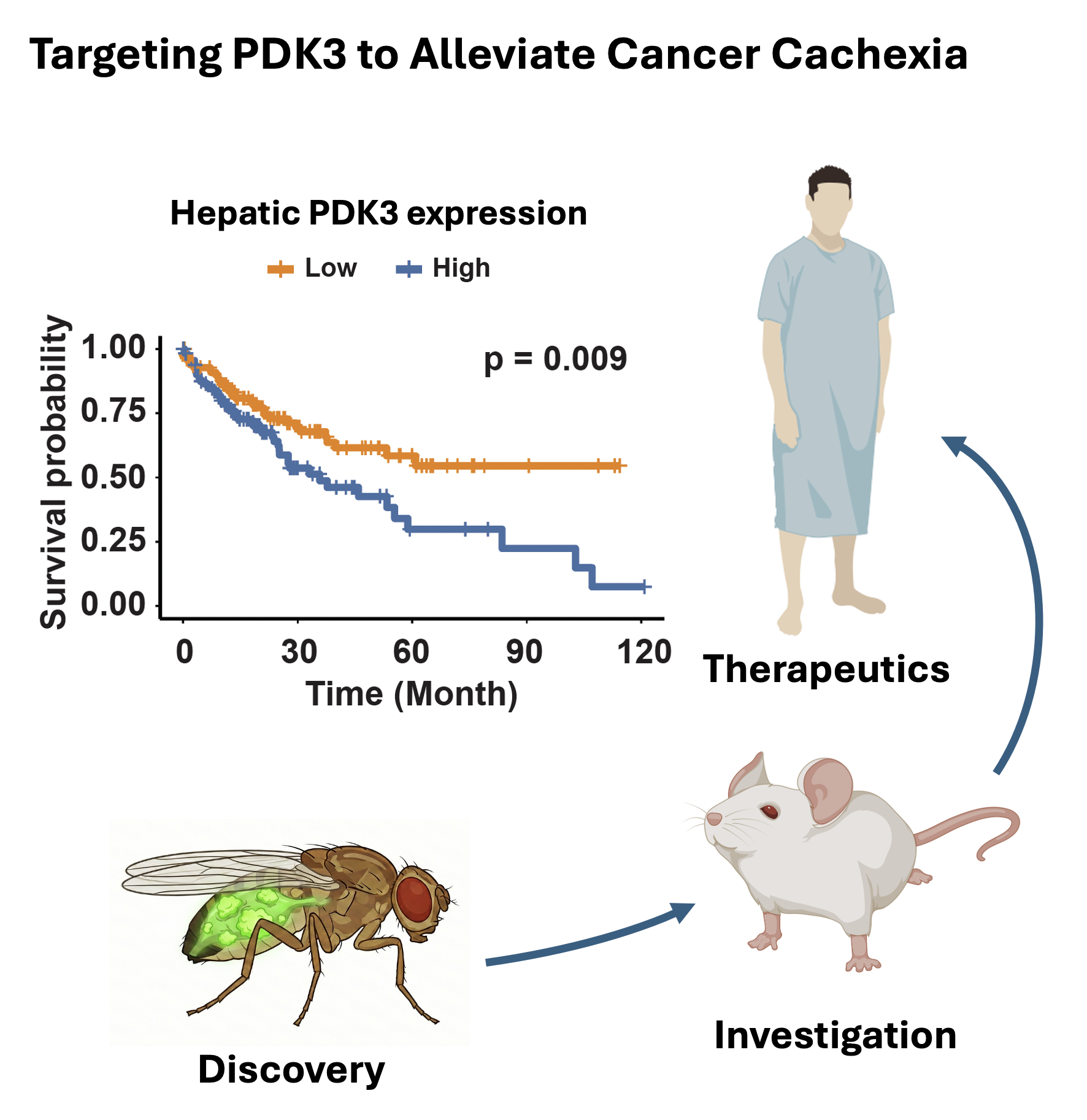
A New Framework for Disease Biology
We study how disease in one part of the body reshapes physiology across the whole organism. By integrating whole-organism biology, evolutionary conservation, and cross-species experimentation, we seek to uncover fundamental mechanisms that drive disease and reveal new opportunities to improve human health.
Step 1
Mapping the Systemic Architecture of Disease
Disease does not respect organ boundaries. We use whole-organism single-cell genomics, genetics, and metabolic profiling to map how local pathology reshapes distant tissues and rewires communication across the body.
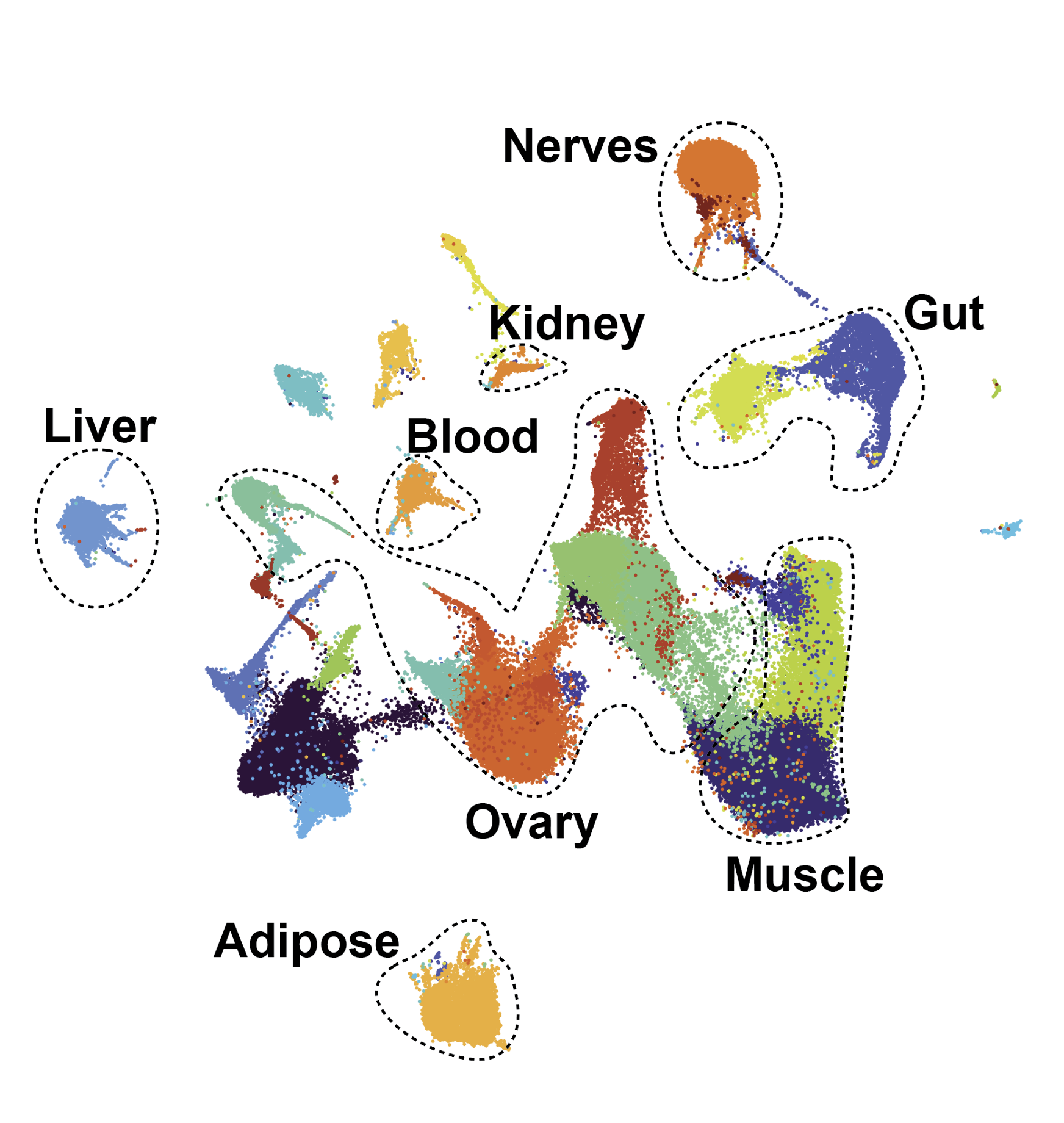
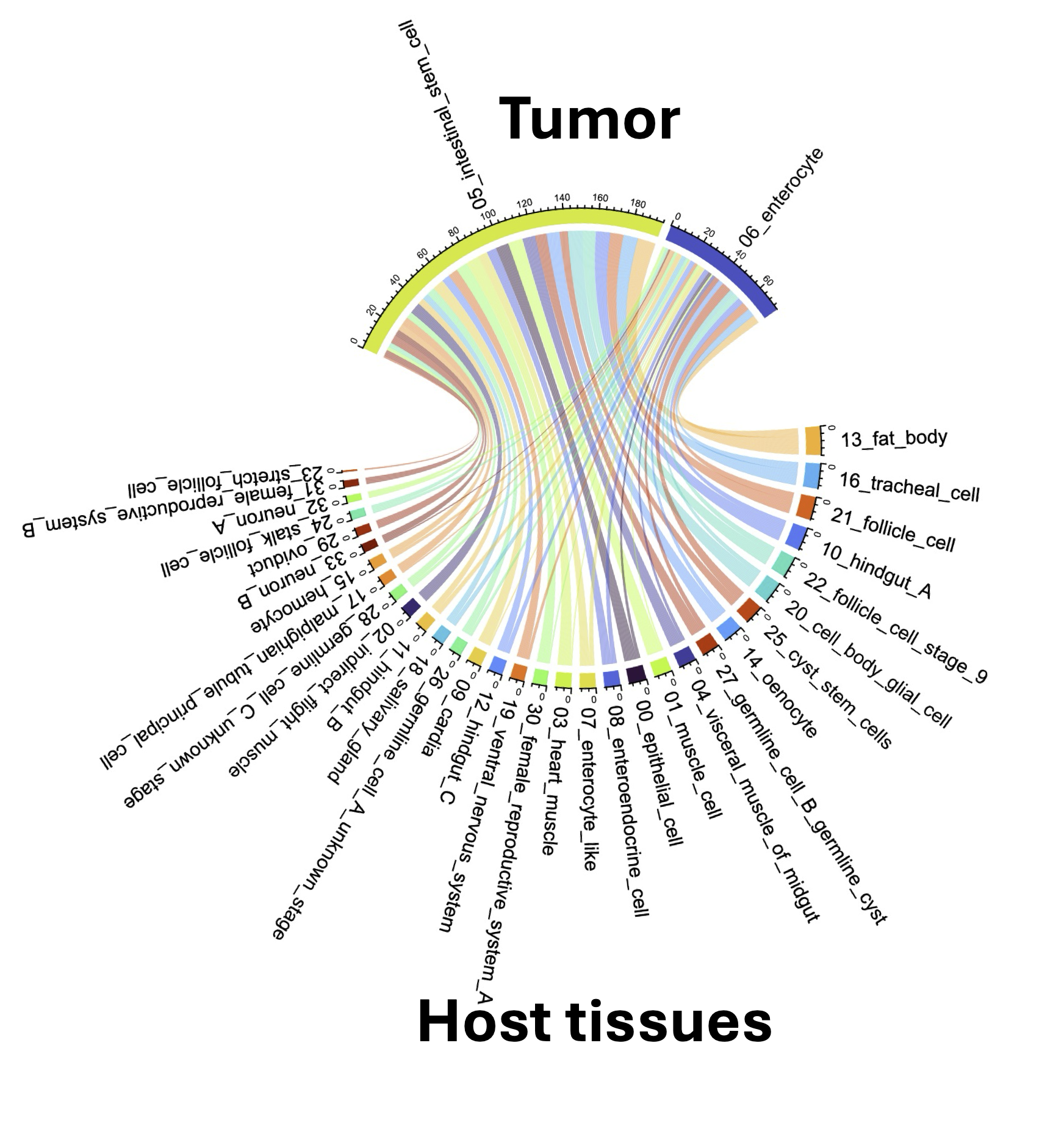
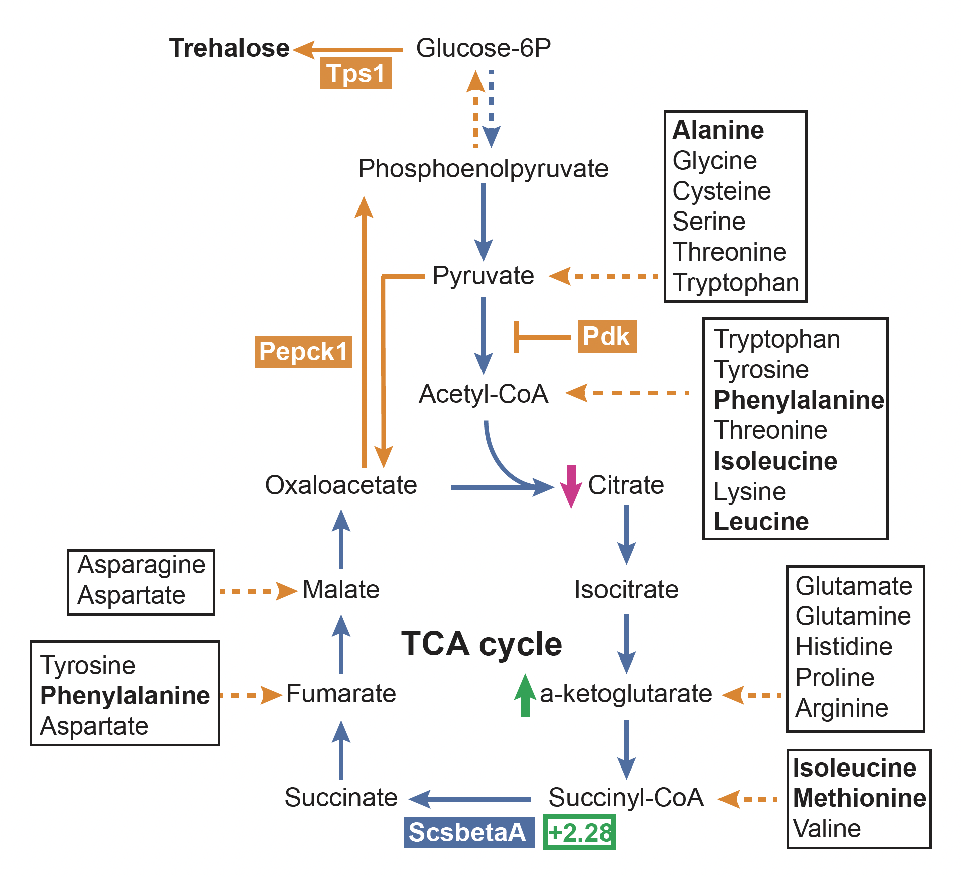
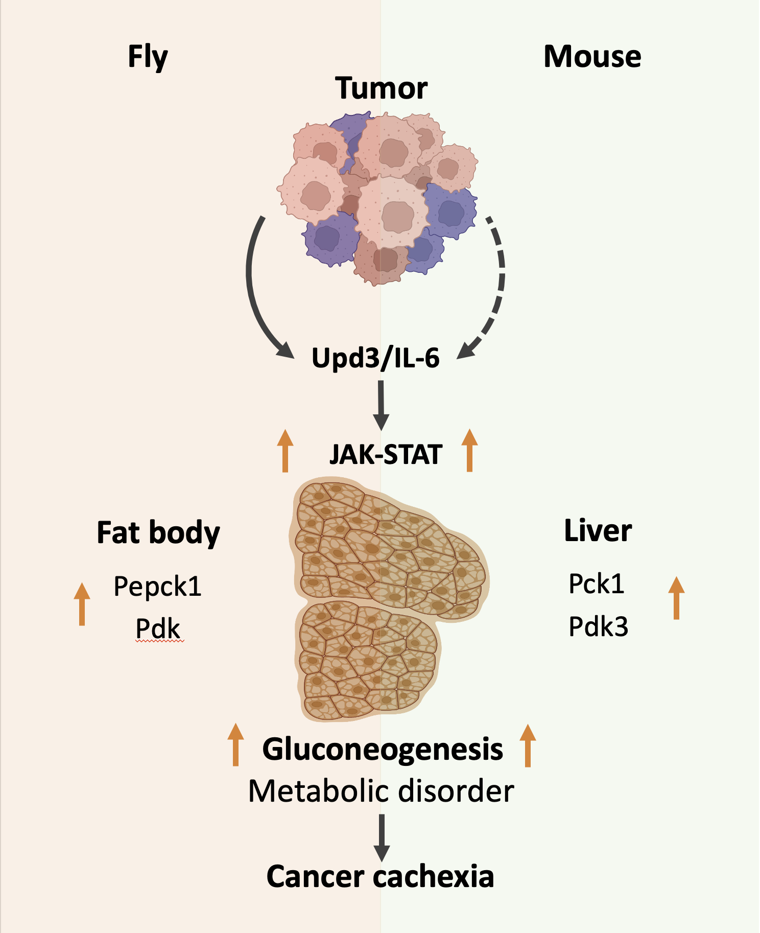
In cancer cachexia, our whole-body single-nucleus studies revealed widespread tumor–host signaling and metabolic reprogramming across multiple organs, creating a systems-level map from which causal mechanisms can be identified and tested.
We do not study organs in isolation—
we study the network that connects them.
Step 2
Evolution as a Discovery Engine
Disease produces thousands of molecular changes, but only a fraction are fundamental drivers.
We use evolutionary conservation as a filter to identify the mechanisms most likely to matter. Rather than asking only whether individual genes are conserved, we compare pathways, cell states, metabolic programs, and inter-organ circuits across species, then test the strongest candidates experimentally for causality.
Evolutionary distance is not a limitation of disease models—it is an experimental variable for discovery.
Step 3
From Mechanism to Human Relevance
Drosophila enables rapid discovery and causal genetics. Mouse models test conserved mechanisms in mammalian physiology. Clinical data and biospecimens establish relevance to human disease.
Embedded within the CSHL–Northwell research ecosystem, the Liu Lab is uniquely positioned to connect these strengths in a bidirectional discovery pipeline. CSHL's NCI-designated Cancer Center provides world-class expertise in fundamental cancer biology and advanced experimental technologies, while Northwell provides access to a large and diverse clinical population, clinicians, biospecimens, and an extensive cancer clinical-trial program. By working across this continuum, we move from mechanistic discovery in tractable models to validation in mammalian and human systems—and bring clinically observed patterns back into the laboratory as experimentally testable questions.